
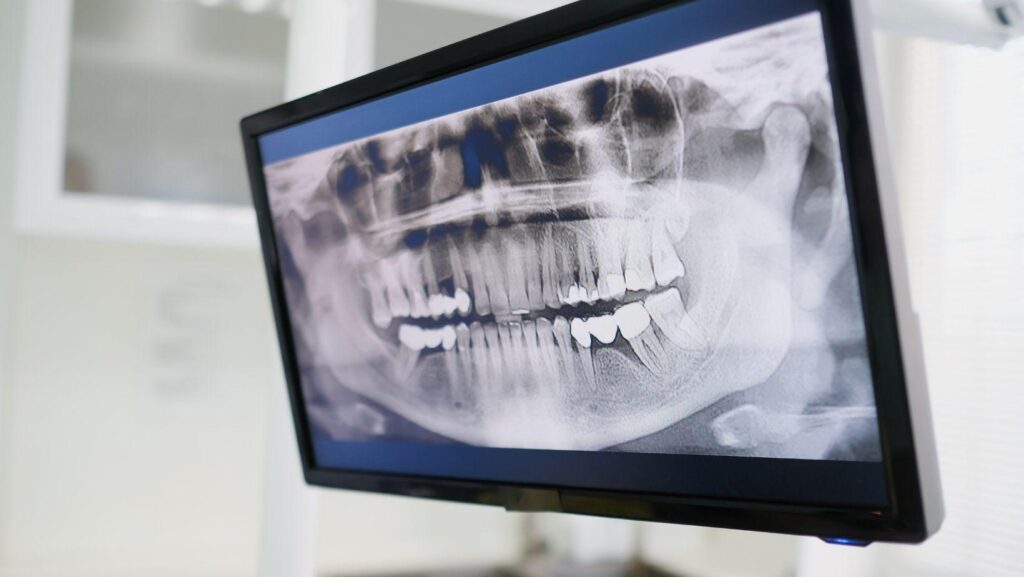
A visual dental exam shows what your dentist can see on the surface, but it cannot reveal every problem beneath your gums or between your teeth. Dental X-rays provide a clearer view of hidden areas, including tooth roots, jawbone, and surrounding structures.
Dental X-rays can detect cavities between teeth, bone loss, root infections, impacted teeth, and other conditions that a visual exam may miss. This kind of detailed imaging is also essential when planning more complex treatment, such as the work done at Florida Dental Implant Center, where X-rays help map bone structure before implant placement. They can also help your dentist assess changes that cause little or no discomfort and use the findings to plan appropriate care.
As you explore how different images support diagnosis, you’ll see why your dentist may recommend X-rays even when your teeth look and feel healthy.
Hidden Problems Below the Gumline
A visual exam cannot show the contact areas between teeth, the bone supporting your teeth, or developing teeth trapped within the jaw. Dental X-rays can reveal these concealed changes before you notice pain, swelling, looseness, or visible damage.
Interproximal Tooth Decay
Decay often starts between neighboring teeth, where toothbrush bristles cannot clean effectively and your dentist cannot see the surfaces directly. Bitewing X-rays can show small areas of mineral loss between teeth, including cavities that look normal during a visual examination.
X-rays may also identify decay beneath fillings, crowns, or other dental work when the surrounding material blocks direct inspection. Your dentist compares the image with your examination findings to determine whether a spot represents active decay, an old restoration, or an area that needs monitoring.
Early detection may allow treatment with a smaller filling instead of waiting until decay reaches the pulp and causes sensitivity or infection.
Periodontal Bone Loss
Gum disease can destroy the bone that supports your teeth without causing noticeable pain. Dental X-rays help your dentist measure changes in the height and shape of the bone around each tooth, especially when gum measurements indicate deeper pockets or bleeding.
The images can show horizontal bone loss, localized defects beside individual teeth, and furcation involvement where bone disappears between the roots of a molar. They may also reveal hardened deposits below the gumline indirectly, although a clinical examination remains necessary to locate calculus.
Your dentist uses the X-ray findings with pocket-depth measurements, bleeding patterns, mobility, and gum recession to assess severity and plan treatment.
Impacted Teeth And Eruption Issues
An impacted tooth cannot emerge normally because another tooth, bone, or soft tissue blocks its path. Panoramic or targeted dental X-rays can show the tooth’s position, angle, root development, and relationship to nearby roots or important jaw structures.
These images commonly help evaluate wisdom teeth, canines that have not erupted, and extra teeth. They can also identify delayed eruption, retained baby teeth, and teeth developing in an abnormal direction.
Your dentist may recommend additional three-dimensional imaging when a tooth lies close to a nerve or when surgery requires more precise detail. A developing problem may remain symptom-free until it causes crowding, root damage, cyst formation, or infection.
Internal Tooth And Root Conditions
A visual exam cannot show the full length of your roots, the pulp chamber, or changes inside a tooth. Dental X-rays can help identify infections, fractures, and treatment-related problems that may not cause obvious symptoms at first.
Root Infections And Abscesses
A periapical X-ray shows the tooth from its crown to the root tip. Your dentist can examine the surrounding bone for a darkened area that may indicate inflammation or an abscess near the root.
These infections often develop after bacteria reach the pulp through deep decay, a damaged filling, or a crack. You may have pain, swelling, sensitivity, or no noticeable symptoms.
An X-ray cannot identify every infection by itself, so your dentist combines the image with an examination, your symptoms, and tests of the tooth’s response. The image can also help your dentist assess whether an infection has affected nearby bone and whether root canal treatment may be appropriate.
Early changes may appear subtle, particularly when the infection has not yet caused significant bone damage.
Cracks And Root Fractures
X-rays may reveal some fractures, but they do not show every crack. A fracture becomes easier to detect when it separates a tooth segment, extends through the root, or causes changes in the surrounding bone.
Your dentist may look for a narrow bone defect alongside the root, a change in the root’s outline, or an infection pattern that does not match ordinary decay. These findings can support a diagnosis when you report sharp pain while biting or discomfort when releasing your bite.
Small cracks in the crown often remain invisible on conventional X-rays. Your dentist may therefore use a bite test, light examination, periodontal probing, or 3D imaging when necessary.
A normal X-ray does not rule out a crack.
Pulp Changes And Prior Treatment Concerns
X-rays let your dentist evaluate the size and shape of the pulp chamber, which contains the tooth’s nerves and blood vessels. Deep decay, repeated dental work, trauma, or aging can cause the chamber to narrow or develop irregular calcifications.
An X-ray also shows the quality of a previous root canal. Your dentist can check whether the filling material reaches the intended length, whether untreated canals may remain, and whether bone changes have developed around the root tip.
These findings help explain persistent pain, tenderness, or swelling after treatment. Because an X-ray shows structure rather than nerve activity, your dentist may also perform temperature, electrical, or tapping tests before deciding on further care.
Jawbone And Surrounding Structure Findings
Dental X-rays can reveal changes beneath your gums, around tooth roots, and within the jawbone that a visual exam cannot show. They may identify cysts, bone loss, sinus changes, impacted teeth, and developmental differences that affect diagnosis or treatment planning.
Cysts And Other Jaw Lesions
X-rays can show cysts, tumors, and other abnormal areas within the jawbone. A cyst may appear as a dark, well-defined space near the root of a tooth or around an unerupted tooth.
Some develop after infection or tooth damage, while others occur near teeth that have not erupted normally. Images can also reveal bone loss from a dental abscess or advanced gum disease.
Radiographs may show whether a lesion has displaced teeth, expanded the bone, or affected nearby structures. These findings do not usually establish a final diagnosis; your dentist may recommend additional imaging, referral, or a biopsy when the appearance is uncertain.
Sinus And Anatomical Changes
X-rays can show the relationship between your upper teeth and the maxillary sinuses, which sit above the back teeth. They may reveal thickened sinus lining, fluid levels, retained roots, or infection-related changes near tooth roots.
These findings can help your dentist distinguish dental causes from conditions that require medical evaluation. Radiographs also display important anatomical structures, including the mandibular canal, mental foramen, nasal cavity, and bone contours.
Knowing their location helps your dentist plan extractions, implants, and other procedures while reducing the risk of injury to nearby nerves or tissues.
Developmental Abnormalities
Dental X-rays can identify teeth that remain impacted, develop in an unusual position, or fail to form. They may reveal extra teeth, missing permanent teeth, delayed eruption, abnormal root shapes, and teeth growing into neighboring roots.
These findings often appear before they cause pain or visible swelling. In children and teenagers, imaging helps your dentist monitor tooth development and detect crowding or eruption problems early.
In adults, it can show retained baby teeth, unerupted wisdom teeth, or structural differences in the jaw. Your dentist selects the imaging type based on the suspected problem; panoramic X-rays provide a broad view, while focused or 3-D scans offer more detail when needed.
Using Imaging To Plan Timely Care
The right image can reveal decay, infection, bone loss, or an impacted tooth that a visual exam cannot show. Your dentist should match the imaging method to your symptoms, examination findings, treatment needs, and prior dental records.
Choosing The Appropriate X-Ray Type
Bitewing X-rays show the crowns of upper and lower teeth together. They help identify decay between teeth and changes near the gumline, including areas that are difficult to inspect directly.
Periapical X-rays capture an entire tooth, including its roots and surrounding bone. Your dentist may use them to investigate tooth pain, an abscess, root damage, or bone changes near a specific tooth.
Panoramic X-rays provide a broad view of your jaws, teeth, and temporomandibular joints. They can help assess developing or impacted teeth, extensive bone changes, and treatment needs that involve multiple areas.
Cone beam computed tomography (CBCT) produces three-dimensional images. Your dentist may recommend it for complex implant planning, oral surgery, unusual root anatomy, or impacted teeth when two-dimensional images do not provide enough detail.
Balancing Diagnostic Value And Radiation Exposure
Your dentist should order an X-ray only when the result could affect diagnosis or treatment. The decision depends on factors such as your age, symptoms, dental history, risk of decay, previous images, and whether a current image already answers the clinical question.
Digital systems generally use less radiation than older film-based systems. Every exposure still requires justification.
Tell your dentist if you are pregnant or might be pregnant. Provide records from previous providers to help prevent unnecessary repeat images.
A CBCT scan can supply valuable three-dimensional information. It usually exposes you to more radiation than a standard dental X-ray.
Your dentist should use the smallest suitable field of view and the lowest exposure that can produce a diagnostically useful image.