
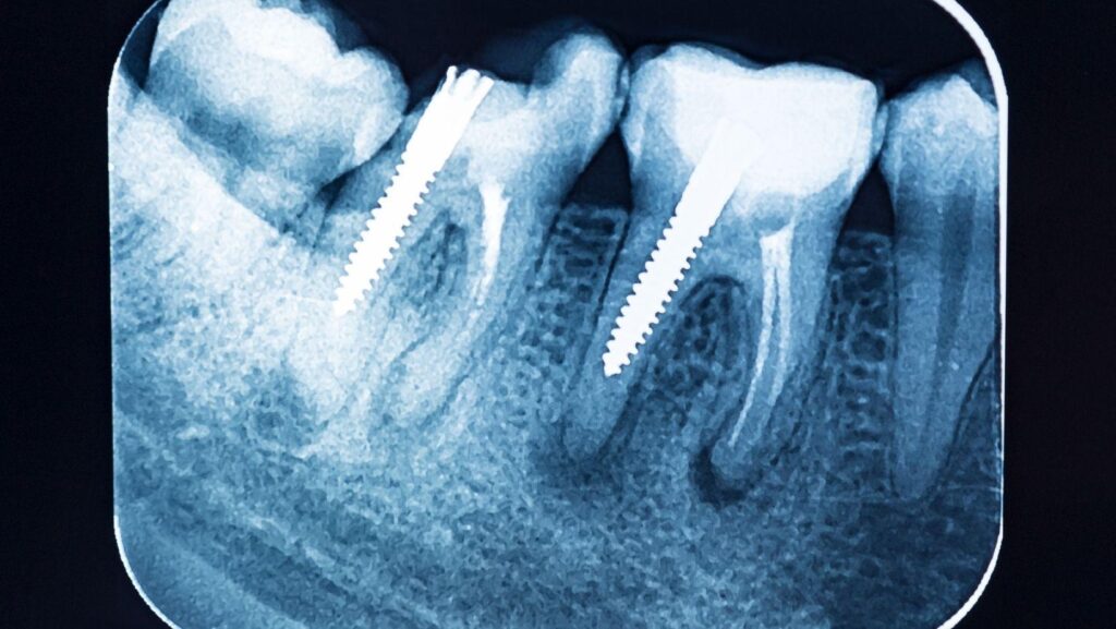
Dental X-rays are part of many dental visits because they show problems that a visual exam cannot detect. Your dentist can use these images to identify cavities between teeth, bone changes, infections, and other concerns before symptoms appear.
You may not need X-rays at every appointment, but your dentist recommends them when your history, exam, age, or oral health makes imaging useful. Practices like RGV Oral & Maxillofacial Surgical Center also rely on advanced imaging to plan more complex surgical treatment when a case is referred to them. The type and timing depend on your individual needs, not a fixed schedule for everyone.
What Dental X-Rays Reveal
Dental X-rays show structures that a visual examination cannot reach, including tooth surfaces between teeth, roots, jawbone, and developing teeth. They help your dentist identify problems early, plan treatment, and compare changes over time.
Hidden Cavities Between Teeth
Small cavities often develop between teeth, where toothbrush bristles and dental instruments cannot see or clean completely. Bitewing X-rays can reveal these areas as darker regions within the enamel or dentin, sometimes before you notice pain or sensitivity.
Your dentist also checks the size and depth of a cavity. A shallow lesion may require fluoride treatment and closer monitoring, while decay that reaches the dentin or pulp may need a filling or root canal treatment.
X-rays can also show recurrent decay beneath existing fillings or crowns. Your dentist must interpret the image alongside your examination and symptoms.
Bone Loss And Gum Disease
X-rays can show the height and density of the bone supporting your teeth. Healthy bone usually forms a consistent pattern around the roots, while gum disease may cause bone levels to drop or appear uneven.
Your dentist compares these findings with gum measurements, bleeding, tooth mobility, and plaque levels. The images can help identify the extent of periodontal damage and guide treatment, such as deep cleaning or referral to a gum specialist.
X-rays do not show gum inflammation directly, so they cannot replace a periodontal examination. They mainly reveal the bone changes that may result from advanced or long-standing gum disease.
Impacted Teeth And Root Problems
X-rays show teeth that remain trapped beneath the gum or inside the jaw. This commonly affects wisdom teeth, but other permanent teeth can also become impacted or develop in an abnormal position.
Your dentist uses the image to assess their angle, depth, and relationship to nearby teeth and nerves. Images also reveal problems around tooth roots, including abscesses, cyst-like lesions, unusual root shapes, and areas of infection or bone destruction.
When a root canal or extraction requires detailed planning, your dentist may recommend a three-dimensional scan rather than a standard dental X-ray. Your dentist chooses the imaging type based on the problem and the treatment you may need.
How Imaging Supports Preventive Care
Dental X-rays reveal structures that a visual exam cannot, including areas between teeth, tooth roots, and supporting bone. They can identify developing problems early and provide images that your dentist can compare during future visits.
Detecting Issues Before Symptoms Begin
You may have tooth decay between teeth or beneath an existing filling before you feel pain. Bitewing X-rays can show these areas, while periapical images can reveal changes near a tooth’s root, such as an infection or bone loss.
Imaging can also help identify impacted teeth, abnormalities in the jaw, and bone changes associated with gum disease. Finding these problems early may allow your dentist to use simpler treatments and prevent damage from progressing.
Your dentist should order X-rays based on your symptoms, examination findings, dental history, and risk factors—not on an automatic schedule. Modern dental imaging uses low radiation doses, but limiting exposure to clinically necessary images remains important.
Tracking Changes Over Time
Comparing current images with earlier X-rays helps your dentist detect changes that may be difficult to recognize during a single examination. For example, repeated images can show whether a suspicious area remains stable, whether decay has enlarged, or whether bone loss is progressing.
Your dentist may use X-rays to monitor developing teeth, evaluate healing after a procedure, or assess the fit and condition of dental work. The timing depends on your age, oral health, symptoms, and treatment needs.
Some patients need imaging more often, while others may safely go longer between scans.
Safety, Frequency, And Types Of Dental X-Rays
Dental X-rays use low radiation doses to reveal problems that a visual examination may miss. Your dentist chooses the imaging type and timing based on your symptoms, oral-health history, age, and risk of decay or gum disease.
Digital Imaging And Radiation Exposure
Digital sensors capture dental images quickly and generally require less radiation than older film systems. Your dental team also uses protective measures, such as positioning equipment carefully and limiting the beam to the area being examined.
Common imaging types serve different purposes:
- Bitewing X-rays: Detect decay between teeth and show bone levels.
- Periapical X-rays: Display an entire tooth, including its root and surrounding bone.
- Panoramic X-rays: Show the jaws, teeth, and developing or impacted teeth in one image.
- CBCT scans: Create detailed three-dimensional views when planning implants, complex extractions, or other treatments.
Dental X-rays are considered safe when your dentist orders them for a clear clinical reason. Tell your dentist if you are pregnant or might be pregnant so the team can consider the timing and necessary precautions.
When Your Dentist Recommends New Images
You may not need X-rays at every appointment. Your dentist reviews your previous images and considers new ones when they could change your diagnosis or treatment.
You might need updated images if you have new tooth pain, swelling, sensitivity, trauma, suspected decay, gum disease, or a failed restoration.
Your dentist may also recommend them before procedures such as root-canal treatment, tooth removal, orthodontic treatment, or implant placement.
The interval varies by risk. A patient with frequent cavities, extensive dental work, dry mouth, or gum disease may need images more often than someone with stable oral health.
Children and teenagers may receive imaging at different intervals because their teeth and jaws continue developing.
Ask which problem an X-ray will evaluate and whether your dentist can use an existing image instead.